3D Cone Beam and 3D Dental Scans Austin, TX
Advancements in dental technology have brought about many new tools that make it easier for dentists and more comfortable for patients. Dental cone beam computed tomography (CBCT), a new type of X-ray equipment, allows dentists to see a clear, detailed, three-dimensional image of the mouth without causing pain to the patient. This 3D imaging system takes full photos of the teeth, soft tissues, nerve pathways, and bones in a single scan.
Cone beam dental scans are available at Austin Lifetime Dental in Austin and the surrounding area. Our staff can help you learn more about the procedure you are undergoing and answer any questions you have about 3D imaging. Call us at (512) 387-9049 to schedule a consultation appointment today.
How 3D Imaging Works
A 3D cone beam machine resembles conventional CT scan machines and comes in two different structures: an upright chair for sitting or a moveable table for lying down. Depending on the procedure and type of machine being used, the patient will be seated in an exam chair or lie down on an exam table. The chair has an extendable arm (C-arm) while the table has a rotator (gantry) that both rotate 360 degrees around the patient's head, taking multiple images at once.
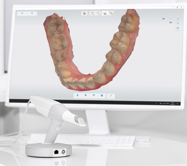
The images are taken at different angles and gathered to create a single 3D image. According to the Radiology organization, "In a single rotation, the detector can generate anywhere between 150 to 200 high resolution two-dimensional (2-D) images, which are then digitally combined to form a 3-D image that can provide your dentist or oral surgeon with valuable information about your oral and craniofacial health." The 3D image is available as soon as the scan is complete, allowing the doctor to discuss the patient's treatment plan in the same visit.
“The chair has an extendable arm (C-arm) while the table has a rotator (gantry) that both rotate 360 degrees around the patient’s head, taking multiple images at once.”
Differences Between Traditional and Cone Beam
Traditional CT scanners and cone beam CT scanners both undertake the same basic function, but technical differences set them apart.
Traditional CT Scans
A traditional CT scan takes several pictures of internal body structures from multiple X-ray images generated by a computer. Atlantis, a radiology equipment company, explains, "The x-rays utilize radiation from a radioactive contrast injected into the body to create cross-sectional images." Traditional CT scans can offer a variety of benefits, primarily for surgeries and diagnostics.
Cone Beam CT Scans
Cone beam scanners use a cone-shaped beam radiating from an X-ray source, covering a wide range with just a single rotation around the patient's head. The X-rays are compiled using a series of algorithms to furnish high-resolution 3D images. Cone beams use a fan beam, as opposed to a light beam, and emit 200-300 times less radiation.
“The X-rays are compiled using a series of algorithms to furnish high-resolution 3D images.”
What a Cone Beam Image Reveals
Cone beam CT scanners reveal far more information than traditional scanners and provide detailed images of a patient's underlying bone structure. Cone beams are primarily used for cases in which traditional X-rays would not provide sufficient information needed for treatments, specifically surgeries and underlying disease. They can evaluate diseases of the jaw, dentition, body structures of the face, nasal cavity, and sinuses.
Cone beam technology has also been useful for diagnosing oral cancers and cysts and managing impacted teeth. A study on cone beam technology found that there has been a "significant contribution of CBCT in the planning and successful surgical management of dentigerous cysts and associated impacted teeth." Since cone beams take wide range photos, they capture in-depth areas that traditional scanners can easily miss.
“They can evaluate diseases of the jaw, dentition, body structures of the face, nasal cavity, and sinuses.”
Check out what others are saying about our dental services on Yelp: 3D Cone Beam and 3D Dental Scans in Austin, TX
What to Expect Post-Exam
The cone beam scan is painless and does not require anesthesia, allowing patients to resume normal activity right after their examination. Although the machine does not cause any pain, claustrophobic should inform their dental practitioner to better accommodate them.
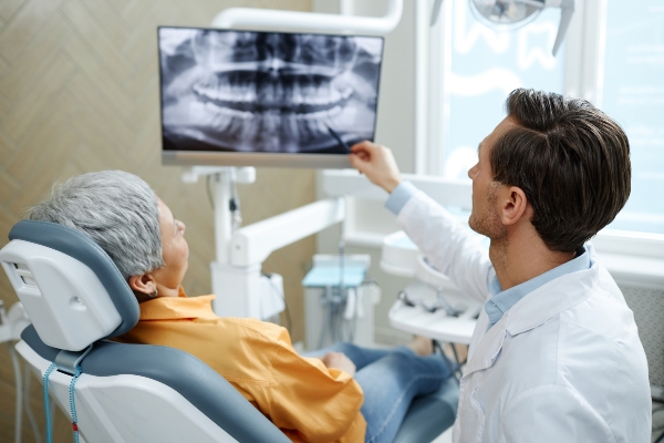
After the exam, the dentist will write a report of the scan results. We will discuss the curated treatment plan with the patient, answering any questions they may have. The patient will be able to see their 3D images and follow the dentist as they move through the treatment plan, pointing at the treatment areas. Preferences for treatments, anesthetics, and medications will also be discussed to fast-track future visits.
“The patient will be able to see their 3D images and follow the dentist as they move through the treatment plan, pointing at the treatment areas.”
Questions Answered on This Page
Q. How does a 3D cone beam work?
Q. What is the difference between traditional and cone beam scanners?
Q. What can a cone beam scan reveal about a patient?
People Also Ask
Q. Who should get tooth fillings?
Q. What should patients do if they have sensitive teeth?
Q. What happens at a dental checkup?
Frequently Asked Questions About 3D Cone Beam Scans
Q. How long do 3D dental scans take?
A. Not very long. A 3D dental scan comprises one rotation of a mechanical arm around your head. After you are positioned, the scan itself usually takes less than 30 seconds.
Q. How long have dentists been using 3D scanning technology?
A. According to the FDA, CBCT technology has been in use for twenty years. Dental scans are becoming more common every day because of their helpfulness in planning for procedures and diagnosing complex conditions.
Q. How much radiation does a 3D scan emit?
A. A 3D dental scanner is considered a computed tomography (CT) scan. Still, it emits less radiation than other conventional CT scans that are commonly used in medicine. However, a CBCT scan does emit more radiation than traditional dental x-rays.
Q. What do patients think of 3D cone beam scanning technology?
A. Patients are often impressed to see a three-dimensional image of their own maxillofacial region. When the dentist can show a patient how a procedure works on their actual mouth and jaw, it helps them have a deeper understanding of the work. Then, patients are more comfortable with the procedure and even have an easier time explaining to others how it works.
Q. What is a 3D cone beam scanner typically used for in dentistry?


Scan here to view this page, 3D Cone Beam and 3D Dental Scans, on mobile
Dental Terminology
Learn More About 3D Scans Today
If you are looking for more information pertaining to complete health dentistry or the use of 3D cone beams or 3D dental scans, call us at 512-387-9049.
Helpful Related Links
- American Dental Association (ADA). Glossary of Dental Clinical Terms. 2025
About our business and website security
- Austin Lifetime Dental was established in 2022.
- We accept the following payment methods: American Express, Cash, Check, Discover, MasterCard, and Visa
- We serve patients from the following counties: Travis County
- We serve patients from the following cities: Austin, West Lake Hills, Barton Creek, Rollingwood, Lost Creek, Tarrytown, Hyde Park, Oak Hill, Austin Lake Hills and Bee Cave
- Norton Safe Web. View Details
- Trend Micro Site Safety Center. View Details

Scan here to open directions to Austin Lifetime Dental on mobile
Back to top of 3D Cone Beam and 3D Dental Scans
Related Posts
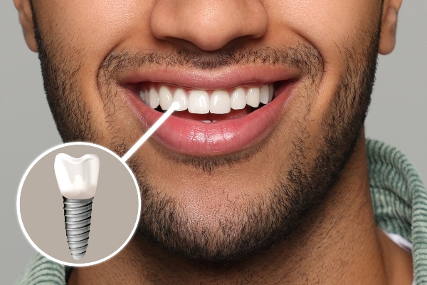
The Ultimate Guide To Receiving Dental Implants
Dental implants can replace all or a few missing teeth. One of their most attractive benefits is that they can last a lifetime with proper care. However, the dental implant process can take longer than some people realize. Here is a breakdown of the process and what patients can expect.Before one can receive dental implants,…
Securely Replace Teeth With Dental Implants
Dental implants are more secure than removable dentures. These restorations can make smiling, speaking, and eating more stable. Embarrassment and awkwardness will be non-existent once these restorations are in place. Here are the details on how dental implants can serve as solid dental replacements.Osseointegration is the process behind the strength and stability of dental implants.…

Exploring Different Types Of Dental Implants And Their Benefits
Dental implants are lasting and stable dental restorations. Research shows that many people prefer implants to removable dentures because of their many benefits. Knowing how implants are better dental replacements can motivate you to come in for a consultation. Here are the benefits of having dental implants as replacements for your missing teeth.Tooth loss results…
Explore additional topics covered on our website:
Call Us Today

Scan here to call Austin Lifetime Dental on mobile